becoming a better Clinician
Nature has located the eyes close to the brain so that its messages may arrive there quickly.
Eyes are fluid filled structures which makes them the perfect organ for ultrasound assessment.
Indications
-
Vision decrease or loss
-
Ocular Pain
-
Foreign body
-
suspected raised Intracranial pressure
-
Eye Trauma
Anatomy
Conjunctiva
-
thin layer of epithelium that protects the cornea
Cornea
-
transparent, thin, hypoechoic
-
responsible for bending light (eg. different bend when underwater but corrects with goggles)
Sclera
-
white part of the eye
-
extrinsic muscles attach here
Choroid
-
network of blood vessels
-
pigmented black
Retina
-
coats the back part of the eye red
-
composed of the photo-receptors
-
reason for "red eye effect" in photography
Ora Serrata
-
transition of the retina from non-photosensitive area to photosensitive area
-
anterior attachment of retina
Fovea
-
dimple in the retina
-
rich in cones
-
in the center of macula
-
posterior attachment of retina
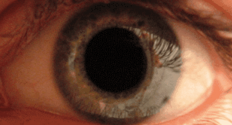
Pupil
-
the black hole (its size is controlled by the iris)
Iris
-
colored part of the eye
-
works much like the diaphragm of a camera by opening and closing
Lens
-
bends light
-
changes shape when zonular fibers via ciliary body contracts/relaxes
Ciliary Body/Muscle
-
changes the shape of the lens when contracts/relaxes
Anterior Chamber
-
filled with anechoic fluid
Aqueous Humor
Vitreous Humor
-
suspends the lens
-
maintains structure of the eyeball
Macula
-
deteriorates with age
-
responsible for the center of our vision
-
macular degeneration patient are left with only peripheral vision
Optic Nerve
-
composed of the retinal axons
-
hypoechoic linear region
-
doppler can be used to assess central retinal artery
-
size: 3mm down (>5 mm suspicious for increased ICP)
Retrobulbar Area
-
includes optic nerve, extraocular muscles and bony orbit


Technique
A high resolution linear probe is used at the highest frequency setting available (>7.5 MHz)
The eye is closed and a small amount of sterile water or large amount of gel is placed on the patient's eye lid. Tegaderm can also be used but make sure there is no air entrapped.
Adjust the depth so that the whole eyeball fills the screen.
The eye is then examined transversely and sagittal (longitudinally), pressing very lightly.
In each transducer position the patient is asked to move the eyes to the left and to the right, and up and down. In this manner the entire eye is examined.
Initially use normal settings, then use very high gain settings to examine the vitreous chamber.
Shining a light into the other eye allows you to see pupilary dilation/constriction on the US screen.

Ruptured Globe

Dislocated Lens

Foreign Body (+ reverberation)